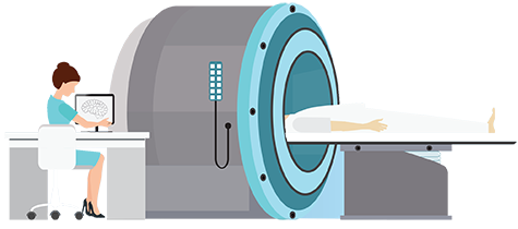

The title “X-ray technician” is a legacy term that predates the 1960s, but the term has persisted in common use. More accurate titles such as “radiographer,” “radiologic technologist” or “X-ray technologist” have been introduced by credentialing and government agencies, including the American Registry of Radiologic Technologists and the Bureau of Labor Statistics.
The change in title reflects both the practical terminology — although the images are commonly referred to as X-rays, the images produced by these X-ray machines are ionizing radiation waves and thus technically radiographs — as well as the evolving responsibility and training of the technologists. The ARRT describes an increased professional responsibility, supported by a “mastery of the principles and practical application of the body of knowledge underlying professional practice.”
While requirements can vary by state, according to the BLS (BLS.gov, 2012) and the ARRT, many states use ARRT exam scores when determining who to license. To sit for the ARRT exam, students must have completed an approved education program. Beginning in 2015, the AART plans to require a postsecondary degree for candidates taking the certification exam. This degree requirement could demonstrate a solid foundation of general education, and a student’s major does not need to be in radiologic sciences.
The American Society of Radiologic Technologists notes that 39 states have licensing requirements for radiography and 32 states have licensing requirements governing limited X-ray machine operators, such a dental assistants or veterinary technicians. Limited X-ray machine operators are defined differently by each state. Some states may define a limited X-ray machine operator as one who works under the supervision of a licensed medical practitioner and who can perform imaging procedures on specified areas, for example:
Limited X-ray machine operators in some states can also work with bone densitometry, which measures the bone density of a patient.
Find a radiology program specializing in X-ray technology by browsing our list of radiology schools or search by radiology degree programs. You can also search X-ray schools by state to find a program offering X-ray technologist courses.
The first radiographs, or X-rays, were taken in 1895 by Wilhelm Roentgen of his wife’s hand, NASA explains. The images clearly showed his wife’s finger bones as well as her wedding ring. Over the next century, radiographic technology has advanced to the point where we can now view highly detailed series of images. The FDA describes these as X-ray “movies,” noting their use in guiding operations or viewing the GI or vascular tracts.
X-rays — specifically, the rays — consist of low-level ionized radiation that passes through the softer parts of the body, such as the skin, muscles and organs, but is blocked by bones and other foreign objects. The images of bones and metal objects are actually shadows on “X-ray film” caused by a lack of X-rays passing through the denser portions of a body.
Some X-ray techniques, such as fluoroscopy, reveal body functions that do not incorporate bones. This is accomplished through the use of a contrast media (a solution that affects X-rays similarly to bones), which is injected or ingested by the person during the procedure. By tracking the media as it flows through the GI tract or through veins or arteries, fluoroscopy can reveal blockages or other ailments that would otherwise only be diagnosed after surgery.
For more information about what opportunities may be available with a degree or certification in radiography, as well as the types of machines technologists and technicians may work with, visit our radiology specializations page.
Depending on the level of certification and licensing, an X-ray technologist can operate the X-ray machine found in dentist offices, clinics and hospital rooms, and can also perform fluoroscopy procedures or operate a computed tomography (CT) machine, which provides X-ray “slices” of the inside of a body or object.According to the BLS (BLS.gov, 2012), X-ray technologists can generally expect to do the following:
Depending on the machine, the x-ray technologist may also prepare the contrast media mixtures for use by the patient. In some cases, this mixture may be radioactive. For more information on some individual qualities that could be useful in this profession, see our X-ray technologist job roles page.
According to the BLS, as of May 2011, the national annual median wage for radiologic technologists and technicians was $55,120, with the lowest 10 percent and highest 10 percent earning $37,360 and $77,760, respectively, during the same period (BLS.gov, 2012). The BLS also reports that, from 2010 to 2020, radiologic technologists and technicians may see an employment growth rate of up to 28 percent, nationally — faster than the average for all occupations (BLS.gov, 2012). Most radiologic technologists are employed by general hospitals or in physicians’ offices.
To see how the salary of radiologic technologist stacks up against other health care professionals, or for salary data for your state, check out our X-ray technologist salary page. Of course, actual salaries can depend on training, experience and geographic location.